
Dental implant surgery in Cambridge utilizes advanced imaging technologies like X-rays and CT scans for precise planning. These non-invasive methods provide detailed, 3D visualizations of the oral cavity, enabling dentists to accurately assess bone density, nerve pathways, and critical structures. By combining these techniques, professionals create personalized treatment plans that enhance implant success rates, preserve natural jaw architecture, and minimize complications. Advanced imaging ensures tailored care for optimal patient outcomes in Cambridge dental implant surgeries.
“Uncovering the foundations of successful dental implant surgery in Cambridge begins with advanced diagnostic imaging. X-rays and CT scans play a pivotal role in planning and executing these complex procedures. This article explores how these techniques, with their remarkable precision, enable dentists to navigate intricate oral anatomies. From identifying bone structures to localizing nerves and blood vessels, modern imaging ensures safe and effective dental implant surgery in Cambridge, enhancing patient outcomes.”
- Understanding Dental Implants: A Foundation for Restorative Dentistry
- The Role of X-rays and CT Scans in Implant Surgery
- Precisely Identifying Bone Structure and Density
- Detecting Nerve and Blood Vessel Locations
- Comparing Diagnostic Images for Comprehensive Planning
- Ensuring Safety and Success with Modern Imaging Techniques
Understanding Dental Implants: A Foundation for Restorative Dentistry
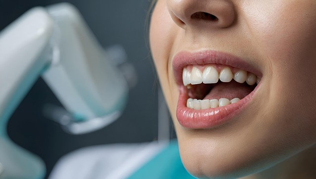
Dental implants represent a cornerstone in restorative dentistry, offering a long-term solution for missing teeth. This advanced procedure involves surgically placing a small titanium post into the jawbone to mimic the natural root structure. Post-surgery, a custom-made dental crown is attached to the implant, providing a secure, functional, and aesthetically pleasing replacement for the lost tooth. For those seeking reliable and durable solutions, dental implant surgery Cambridge has become a popular choice, leveraging cutting-edge technology and expert oral surgeons to restore smile aesthetics and oral function.
Unlike traditional dentures or bridges, implants are designed to fuse with the jawbone, ensuring stability over time. This fusion also helps preserve bone density, a significant concern after tooth loss. With proper care, dental implants can last for decades, making them a smart investment in long-term oral health. Advanced imaging techniques like X-rays and CT scans play a crucial role in planning and executing this precise surgery by providing detailed insights into jaw structure and bone density, essential for ensuring the success of dental implant surgery Cambridge.
The Role of X-rays and CT Scans in Implant Surgery

Dental implant surgery in Cambridge often relies on advanced imaging techniques like X-rays and CT scans to ensure precise planning and successful outcomes. These technologies provide detailed, high-resolution images of the jawbone, teeth, and surrounding structures. For dental implants, X-rays help identify bone density, detect any abnormalities or diseases within the bone, and assess the overall health of the jaw.
CT scans, on the other hand, offer a 3D view, allowing dentists to evaluate the anatomical structure in greater detail. This is particularly crucial for placing implants accurately. By combining these imaging methods, dental professionals in Cambridge can create personalized treatment plans, ensuring that each implant is strategically placed and aligned for long-term success, enhancing the overall success rate of dental implant surgery.
Precisely Identifying Bone Structure and Density

Advanced imaging technologies like X-rays and CT scans play a pivotal role in precisely identifying bone structure and density, especially when considering dental implant surgeries in Cambridge. These non-invasive procedures provide detailed visual data, enabling dentists to accurately assess the health and strength of bones. By examining the density and integrity of the jawbone, professionals can make informed decisions about implant placement, ensuring optimal support for artificial teeth.
In the context of dental implant surgery Cambridge, CT scans offer a three-dimensional view, allowing specialists to map out precise trajectories for implants. This level of detail is crucial in maintaining the natural architecture of the jaw and promoting successful osseointegration—the fusion of the implant with bone tissue. Accurate identification of bone structure also helps in avoiding critical structures like nerves and blood vessels, minimising potential complications during surgery.
Detecting Nerve and Blood Vessel Locations

When it comes to dental implant surgeries in Cambridge, precise localization of nerves and blood vessels is paramount for a successful procedure. Advanced imaging techniques like X-rays and CT scans play a pivotal role in achieving this goal. These technologies offer detailed, three-dimensional visualizations of the oral cavity, allowing dental professionals to accurately identify and map critical anatomical structures.
By analyzing X-ray images, dentists can pinpoint the exact positions of teeth, bone density, and surrounding nerves. CT scans further enhance this process by providing cross-sectional images, enabling precise detection of blood vessels, sinuses, and other vital structures. This detailed information is crucial for planning the dental implant placement, ensuring minimal disruption to surrounding tissues and enhancing the overall success rate of the surgery.
Comparing Diagnostic Images for Comprehensive Planning

When considering dental implant surgery in Cambridge, comparing diagnostic images such as X-rays and CT scans is crucial for comprehensive planning. These advanced imaging techniques provide detailed insights into jawbone density, nerve pathways, and surrounding structures, enabling dentists to make informed decisions about the feasibility and optimal placement of implants.
X-rays offer a straightforward view of dental health, while CT scans deliver three-dimensional models, enhancing accuracy. By integrating these diagnostic tools, oral surgeons in Cambridge can tailor treatment plans, ensuring the successful integration of dental implants while minimising potential complications. This meticulous approach maximises patient outcomes and increases the chances of a long-lasting, functional, and aesthetically pleasing restoration.
Ensuring Safety and Success with Modern Imaging Techniques

At our dental clinic in Cambridge, we prioritize patient safety and comfort when it comes to diagnostic imaging. Modern techniques like X-rays and CT scans play a pivotal role in diagnosing complex oral issues accurately, especially before undergoing procedures like dental implant surgery. These advanced technologies offer precise, detailed images of the mouth’s internal structures, enabling our skilled dentists to identify problems that might not be apparent through visual examination alone.
By utilizing modern imaging techniques, we ensure successful outcomes for all treatments, from routine check-ups to complex reconstructive surgeries. Our state-of-the-art equipment is regularly maintained and calibrated to deliver consistent, high-quality images. This commitment to excellence in diagnostics translates into better patient care, allowing us to tailor treatment plans that address specific needs, ensuring the best possible outcomes for dental implant surgery Cambridge patients.
Dental implant surgery in Cambridge relies heavily on advanced imaging techniques like X-rays and CT scans, which play a pivotal role in diagnosing and planning. These tools enable dentists to precisely map bone structures, identify nerve and blood vessel locations, and compare diagnostic images for comprehensive treatment planning. Modern imaging ensures safety and success by providing accurate, detailed insights into the patient’s oral health, making dental implant surgery more effective and less invasive than ever before.